

What causes arterial ulcers, and how do they differ from venous ulcers?
Accurately distinguishing arterial ulcers from venous ulcers is a critical clinical safety priority. Appropriate wound care can consist of applying high-compression wraps, a standard therapy for venous ulcers, to an arterial ulcer can completely block remaining arterial blood supply, leading to rapid tissue death and limb loss.
Arterial ulcers, also known as ischemic ulcers, are chronic, non-healing wounds caused by severe impairment of arterial blood flow (ischemia) to the lower extremities, most commonly stemming from Peripheral Artery Disease (PAD) or atherosclerosis.
When narrowed or occluded arteries fail to deliver adequate oxygenated blood and nutrients to distal tissues, cellular hypoxia occurs, leading to tissue necrosis and skin breakdown. In contrast, venous ulcers result from failing vein valves and fluid pooling (venous stasis), producing distinctly different physical symptoms, fluid levels, and clinical management requirements.
| Clinical Feature | Arterial Ischemic Ulcers | Venous Stasis Ulcers |
|---|---|---|
| Primary Underlying Etiology | Arterial occlusion, atherosclerosis, or Peripheral Artery Disease (PAD)[cite: 24] | Venous valve incompetence, deep vein thrombosis (DVT), chronic venous hypertension[cite: 24] |
| Anatomical Wound Location | Distal extremities: toes, heels, lateral malleolus, pressure points of feet[cite: 24] | Medial gaiter region of lower leg (above medial ankle bone)[cite: 24] |
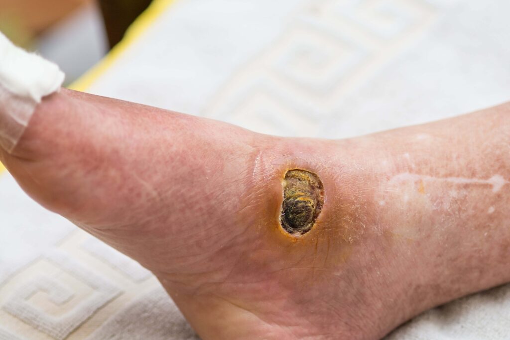
| Visual Wound Characteristics | “Punched-out” round margins, pale or yellow wound bed, dry eschar[cite: 24] | Shallow, irregular margins, ruddy beefy-red granulation tissue, yellow slough[cite: 24] |
| Exudate / Drainage Level | Minimal to zero drainage; dry, desiccated tissue bed[cite: 24] | Moderate to heavy, copiously draining serosanguinous fluid[cite: 24] |
| Surrounding Skin & Limb Signs | Pale, shiny, hairless, cold skin; thick nails; faint or absent peripheral pulses[cite: 24] | Hyperpigmented (hemosiderin staining), warm skin; lower limb edema (swelling)[cite: 24] |
| Pain Characteristics | Severe, sharp, nocturnal pain elevated by leg elevation; relieved by dangling legs[cite: 24] | Dull, aching, heavy pain that improves when legs are elevated above heart level[cite: 24] |
What are the primary risk factors for developing arterial ulcers?
The primary risk factors for developing arterial ulcers include systemic vascular conditions that damage arterial walls or promote plaque buildup, such as uncontrolled Diabetes Mellitus, active tobacco smoking or nicotine use, severe hypertension, hyperlipidemia, and advanced age. These metabolic and lifestyle factors accelerate the progression of Peripheral Artery Disease (PAD), steadily restricting microvascular lumen diameter until minor pressure or skin friction triggers tissue necrosis.
Managing systemic risk factors is an essential component of long-term arterial ulcer prevention and wound healing protocols.
Systemic Risk Factors
- Tobacco Smoking and Nicotine Use: Nicotine causes immediate peripheral vasoconstriction while tobacco toxins induce chronic vascular endothelial inflammation, drastically accelerating plaque formation.
- Diabetes Mellitus: Chronic high blood sugar damages peripheral microvasculature (microangiopathy) while causing peripheral neuropathy, preventing patients from feeling minor foot trauma.
- Hyperlipidemia and Atherosclerosis: High levels of low-density lipoprotein (LDL) cholesterol form rigid fibrous plaques within arterial walls, narrowing blood vessel pathways.
- Hypertension (High Blood Pressure): Chronic arterial pressure causes vessel wall thickening and arterial stiffness, reducing capillary perfusion capacity.
- Advanced Age and Sedentary Lifestyle: Natural vascular compliance decreases with age, while lack of exercise reduces collateral circulation development.
What non-invasive diagnostic tests are used to evaluate arterial circulation?
Clinicians use non-invasive diagnostic tests such as the Ankle-Brachial Index (ABI), Toe-Brachial Index (TBI), Transcutaneous Oxygen Pressure (TcPO2) monitoring, and arterial Duplex ultrasound to quantify lower extremity blood flow before initiating wound treatment. The Ankle-Brachial Index compares systolic blood pressure measured at the ankle to that of the arm, providing a numerical ratio that reflects the severity of peripheral arterial blockade.
Performing these diagnostic checks is mandatory prior to applying compression wraps or conducting sharp wound debridement.
ABI Score > 1.30: Non-Compressible Calcified Vessels (Common in Severe Diabetes)
│
ABI Score 0.90 – 1.30: Normal Arterial Blood Perfusion
│
ABI Score 0.70 – 0.89: Mild Peripheral Artery Disease (PAD)
│
ABI Score 0.50 – 0.69: Moderate PAD (Compromised Healing Capacity)
│
ABI Score < 0.50: Severe PAD / Critical Limb Ischemia (High Amputation Risk)
Diagnostic Assessment Breakdown
- Ankle-Brachial Index (ABI): An ABI value between 0.90 and 1.30 is normal. An ABI below 0.50 indicates severe arterial obstruction and Critical Limb Ischemia (CLI), requiring immediate vascular surgical referral.
- Toe-Brachial Index (TBI): Used when ankle arteries are rigid and non-compressible due to medial arterial calcification (common in diabetic patients). A TBI below 0.70 indicates significant arterial disease.
- Transcutaneous Oxygen Pressure (TcPO2): Measures the amount of oxygen actually reaching the dermal microvasculature. Values below 30 mmHg signal inadequate oxygenation for spontaneous wound healing.
- Arterial Duplex Ultrasound & Angiography: Visualizes arterial blood flow velocity and maps anatomical sites of arterial narrowing or occlusion.
How are arterial ulcers medical treated and managed?
Medical management of arterial ulcers focuses on restoring arterial blood supply (revascularization), protecting the dry wound bed from infection, managing localized pain, and avoiding inappropriate compression or aggressive debridement. Revascularization procedures performed by a vascular surgeon—such as balloon angioplasty, stenting, or arterial bypass surgery—are frequently required before the wound can receive enough oxygen to regenerate tissue.
Unlike moist wound healing protocols for other chronic sores, stable, dry, intact eschar on an ischemic foot or heel must be kept dry and protected until vascularity is established.
Core Wound Care Strategies
- Vascular Surgical Revascularization: Re-opening blocked arteries using minimally invasive endovascular balloon angioplasty/stenting or surgical bypass grafting to restore tissue oxygenation.
- Dry Protective Wound Dressings: Applying dry sterile gauze, povidone-iodine (Betadine) paint, or non-adherent protective covers to dry, intact eschar to keep bacterial counts low and prevent moisture buildup.
- Avoidance of High Compression: Strict avoidance of multi-layer compression wraps or tight elastic bandages, which cut off remaining fragile arterial blood flow.
- Pharmacological Therapy: Prescribing antiplatelet agents (e.g., aspirin or clopidogrel) and hemorheologic agents (e.g., cilostazol) to improve red blood cell flexibility and microvascular flow.
- Pain Management & Dependent Positioning: Managing severe nocturnal ischemic rest pain by instructing patients to avoid leg elevation and keep affected limbs in a dependent (dangling) position to assist blood flow via gravity.
What is the role of debridement in arterial ulcer care, and when is it contraindicated?
Debridement plays a selective role in arterial ulcer care: it is strictly contraindicated for stable, dry, intact eschar on ischemic limbs without adequate blood flow, but becomes mandatory if the eschar becomes soft, infected, or purulent (unstable). Performing sharp debridement on an ischemic wound without sufficient arterial circulation exposes underlying deep tissue or bone to environmental pathogens that the body cannot fight due to lack of blood-borne immune cells.
A vascular assessment (such as ABI or TBI) must always precede debridement to prevent converting a stable dry lesion into an open, gangrenous wound.
Indications vs. Contraindications for Debridement
- CONTRAINDICATED (Do NOT Debride): Hard, dry, dark, firmly attached eschar on heels, toes, or feet with an ABI below 0.50 or absent pedal pulses, displaying no signs of infection (no redness, warmth, drainage, or odor). The eschar acts as a natural, sterile cover.
- MANDATORY (Must Debride): Unstable, soft, boggy, or fluctuating tissue beneath eschar, accompanied by purulent drainage, foul odor, surrounding cellulitis, or systemic fever. Immediate surgical debridement is necessary to drain underlying infection and prevent sepsis.
- SAFE TO DEBRIDE: Clean, non-viable tissue in a wound that has successfully undergone revascularization and has documented adequate arterial blood supply (ABI above 0.70 or TcPO2 above 30 mmHg).
How do mobile wound care services help manage arterial ulcers at home?
For elderly or immobile individuals suffering from severe arterial pain and walking limitations, mobile care eliminates the physical trauma of clinic travel while ensuring high-risk ischemic wounds receive continuous specialist monitoring.
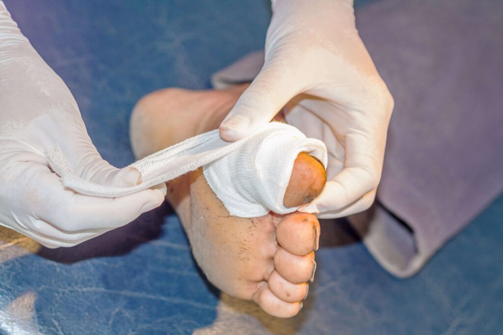
Mobile wound care services assist patients with arterial ulcers by delivering specialized bedside vascular assessments, non-invasive diagnostic testing, protective dressing changes, and vascular surgical coordination directly to the patient’s home, assisted living community, or skilled nursing center.
Managing arterial ulcers requires balancing infection prevention, vascular testing, and surgical referrals. Mobile wound care teams deliver this complex care directly to the patient’s bedside.
Benefits of In-Home Mobile Care for Arterial Ulcers
- In-Home Non-Invasive Vascular Assessment: Licensed wound care specialists perform bedside Doppler pulse checks, Ankle-Brachial Index (ABI) measurements, and tissue oxygenation evaluations without requiring hospital visits.
- Bedside Moisture Control & Protective Dressing Changes: Mobile teams monitor dry eschar, apply protective antimicrobial paints (e.g., povidone-iodine), and adjust non-adherent dressings to keep stable wounds safe.
- Direct Coordination with Vascular Surgeons: When diagnostic checks reveal severe ischemia (ABI below 0.50), mobile providers immediately initiate urgent referrals to vascular surgeons for revascularization procedures.
- Early Infection Detection & Bedside Management: Routine home visits allow clinicians to catch early signs of sub-eschar infection, boggy tissue breakdown, or cellulitis, prescribing targeted antibiotics before gangrene develops.
- Reduced Transport Pain & Amputation Risk: Keeps fragile, painful lower extremities rested at home, preventing friction trauma during transport while dramatically lowering amputation and hospital readmission rates.