
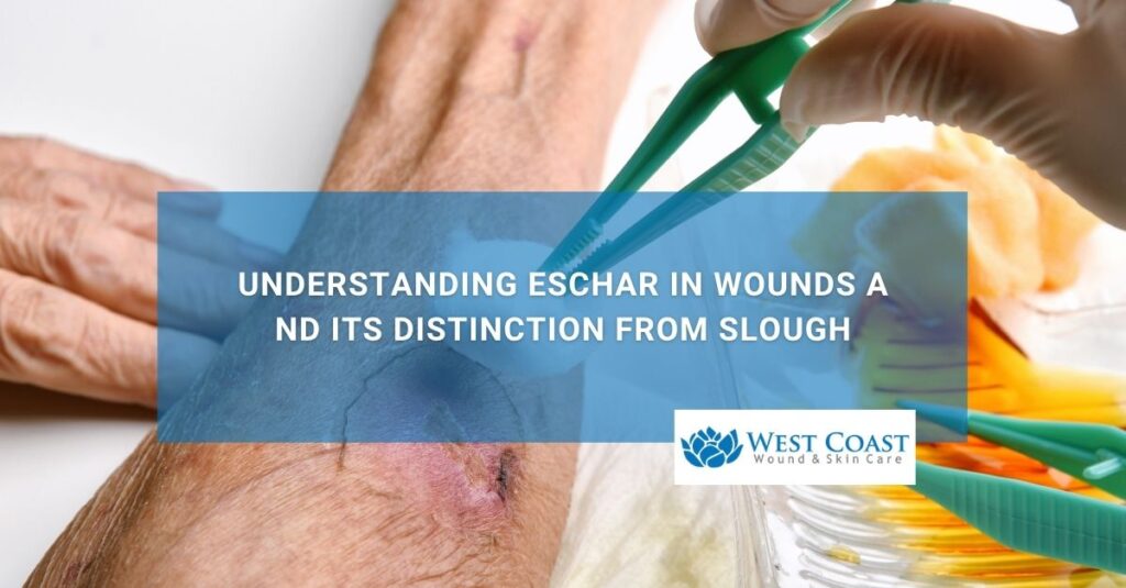
What is eschar in a wound?
Eschar is a thick, dry, leathery layer of dead (necrotic) tissue that forms over deep full-thickness wounds, pressure injuries, severe burns, or arterial ulcers.The true depth of tissue damage, involvement of underlying anatomical structures (such as fascia, tendon, or bone), and overall wound stage cannot be determined until the non-viable tissue is debrided or cleared. Only then is wound care possible.
In clinical staging, particularly with pressure injuries, when a wound bed is completely covered by eschar or slough, clinicians categorize it as Unstageable.
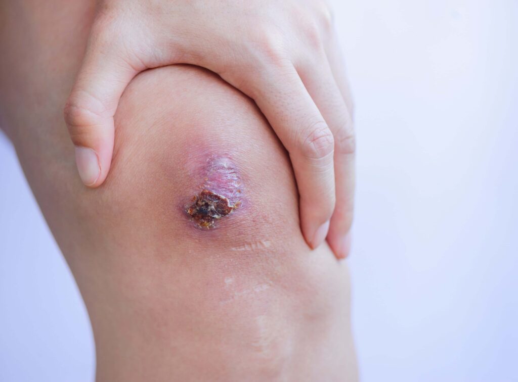
Eschar typically ranges in color from dark tan and brown to coal black and adheres tightly to the underlying wound bed. Because eschar obstructs visual clinical inspection and physically blocks healthy tissue regeneration, specialized evaluation by a clinical wound care team is required to assess its stability and determine the appropriate treatment plan.
Unlike minor superficial crusts or natural scab formations, eschar is a manifestation of significant vascular impairment, deep thermal injury, or severe mechanical pressure that has led to full-thickness tissue necrosis. The dead tissue consists of denatured proteins, dried blood, dead skin cells, and cellular debris trapped within the dermal and subcutaneous layers.
Common Clinical Causes and Types of Wounds Exhibiting Eschar
- Stage 3 and Stage 4 Pressure Injuries (Bedsores): Prolonged unrelieved pressure over bony prominences—such as the sacrum, ischial tuberosity, hips, and heels—restricts capillary blood flow, leading to localized deep-tissue ischemic necrosis and eschar formation.
- Diabetic Foot Ulcers (DFUs): A combination of peripheral neuropathy, microvascular damage, and impaired immune response in diabetic patients often leads to unperceived trauma, tissue necrosis, and dark, firmly attached eschar.
- Arterial and Ischemic Ulcers: Peripheral Artery Disease (PAD) and severely compromised arterial circulation prevent oxygen-rich blood from reaching the lower extremities, causing localized tissue death that hardens into dry eschar.
- Full-Thickness (Third-Degree) Burns: Thermal, chemical, or electrical injuries destroy all layers of the skin, converting the damaged dermis into a rigid, non-viable eschar shell.
- Traumatic Wounds and Surgical Dehiscence: Severe crush injuries, surgical wound breakdown, or compromised skin flaps can develop necrotic eschar if blood supply is disrupted during the inflammatory phase of healing.
What is the difference between eschar, slough, and a scab?
While eschar, slough, and scabs are all non-viable tissue structures that form on damaged skin, they differ significantly in moisture content, biological composition, structural depth, and clinical management requirements. A scab is a temporary, superficial biological crust that forms over minor cuts or abrasions.
In contrast, eschar is a hard, black or dark brown leathery plaque resulting from deep tissue death, while slough is a soft, moist, yellow, tan, or white fibrous debris composed of fibrin, pus, and dead cellular material.
Accurately distinguishing between these three tissue types is critical because applying the wrong wound care protocol can delay healing, trap bacterial bioburden, or inadvertently cause severe tissue damage.
| Clinical Feature | Eschar | Slough | Scab |
|---|---|---|---|
| Visual Appearance | Dark brown, tan, or coal black; dry, firm, rigid, and leathery[cite: 20] | Pale yellow, white, tan, or green; soft, moist, stringy, or mucinous[cite: 20] | Light red, reddish-brown, or dark crust; dry and rough surface[cite: 20] |
| Tissue Depth & Origin | Full-thickness dermal or subcutaneous tissue death (necrosis)[cite: 20] | Inflammatory exudate, accumulated fibrin, white blood cells, and cellular debris[cite: 20] | Superficial epidermal damage with dried blood plasma and platelets[cite: 20] |
| Adhesion to Wound Bed | Extremely tight; strongly bonded to underlying tissue[cite: 20] | Variable; ranges from loosely attached strands to adherent sheets[cite: 20] | Surface level; loosely attached to upper epidermal margins[cite: 20] |
| Primary Clinical Goal | Clinical assessment for debridement vs. dry maintenance[cite: 20] | Systematic removal to reduce bacterial bioburden and exudate[cite: 20] | Protect wound bed and allow natural shedding as epidermis repairs[cite: 20] |
| Common Associated Conditions | Deep pressure ulcers, arterial insufficiency, third-degree burns[cite: 20] | Chronic venous stasis ulcers, delayed-healing surgical wounds[cite: 20] | Minor scrapes, superficial cuts, shallow abrasions, bug bites[cite: 20] |
Biological Differences and Healing Roles
- Scabs: Form as part of the body’s natural hemostatic response to superficial epidermal trauma. Platelets and fibrin trap red blood cells to form a dry temporary protective cap. As new epidermal cells migrate across the wound bed underneath, the scab naturally loosens and falls off without clinical intervention.
- Slough: Represents a continuous inflammatory state in chronic, non-healing wounds. As white blood cells attempt to break down damaged tissue, cellular debris and fibrin accumulate, creating a moist, protein-rich environment. If left untreated, slough creates an ideal breeding ground for bacterial biofilms and polymicrobial infection.
- Eschar: Represents an entire section of dead, devitalized tissue. Because it lacks blood flow, immune cells cannot penetrate it, making it vulnerable to bacterial colonization under its surface if moisture becomes trapped underneath.
Does eschar always need to be removed or debrided for wound care?
No, eschar does not always require immediate clinical removal or debridement. While debridement is standard procedure for most chronic wounds to expose healthy tissue and clear infectious material, stable, dry, intact eschar on ischemic limbs or heels without surrounding redness (erythema), swelling, or drainage should generally be left intact.
In these specific vascular scenarios, the hard eschar serves as the body’s natural, sterile biological cover while circulation is evaluated or re-established.
Determining whether to debride or preserve eschar requires a detailed clinical vascular assessment conducted by a qualified wound care practitioner or mobile specialist team.
When to Preserve Eschar (Leave Intact)
- Stable, Dry Heel Eschar: If a patient has a dry, hard, dark eschar on the heel or lower extremity that exhibits no signs of softness (fluctuance), tenderness, drainage, odor, or surrounding skin redness, it should not be debrided. The heel has limited tissue depth separating skin from bone (calcaneus), and debriding stable eschar in a limb with poor arterial blood flow can expose bone to severe infection or osteomyelitis.
- Severe Peripheral Artery Disease (PAD): When a patient lacks adequate blood perfusion to heal a wound, removing eschar exposes underlying tissue that cannot receive enough oxygen and nutrients to regenerate. The eschar is left dry and intact until a vascular surgeon can evaluate options to restore circulation.
When to Debride Eschar (Mandatory Removal)
- Signs of Infection or Sub-Eschar Abscess: If the eschar becomes soft, boggy, or fluctuating, or if there is visible pus, foul odor, surrounding erythema, warmth, or systemic fever, the eschar must be urgently debrided to drain underlying bacterial infection.
- Active Healing Goals with Adequate Perfusion: When vascular testing confirms sufficient blood flow to support tissue regeneration, removing eschar exposes the viable wound bed, allowing granulating tissue to form and enabling advanced cellular therapies to work.
Overview of Clinical Debridement Methods
- Sharp / Surgical Debridement: The rapid, precise removal of non-viable tissue using sterile scalpels, scissors, or curettes. Performed by licensed clinicians, this is the fastest method to convert an unstageable wound into a clean, healing environment.
- Enzymatic Debridement: The topical application of prescriptive collagenase ointments that specifically break down the collagen strands holding necrotic tissue to the wound bed. This is ideal for patients who cannot tolerate sharp debridement.
- Autolytic Debridement: Utilizing moisture-retentive wound dressings (such as hydrogels or hydrocolloids) to trap the body’s natural enzymes, allowing them to slowly liquefy dry eschar over time.
- Mechanical Debridement: Physical removal of dead tissue using specialized wound irrigation systems, wet-to-dry dressings, or low-frequency ultrasound therapy.
What are the best wound dressings for treating wounds with eschar?
The best dressing for a wound with eschar depends entirely on the overarching clinical objective: whether the goal is to soften and break down dry eschar through autolytic debridement or to keep the eschar dry, protected, and stable.
Moisture-donating dressings such as hydrogels are preferred when hydrating and removing eschar, while protective dry dressings or povidone-iodine preparations are used when keeping stable eschar dry on ischemic limbs.
Selecting an inappropriate dressing can cause serious complications—such as turning a dry, stable eschar into a moist, infected wound bed.
Dressings for Softening and Removing Eschar (Autolytic Debridement)
- Hydrogel Dressings: Formulated with high water content, hydrogels donate moisture to dry, hard eschar, softening the necrotic tissue and allowing native enzymes to break down denatured proteins. They are typically covered with a secondary moisture-retentive dressing.
- Hydrocolloid Dressings: These occlusive, moisture-retentive dressings trap natural wound fluid against the eschar, creating a warm, moist environment that accelerates autolysis while shielding the area from external fluid and bacterial contaminants.
- Medical-Grade Honey (Leptospermum): Provides osmotic action that draws lymph fluid from deeper tissues to hydrate dry eschar while delivering natural antimicrobial activity to control bioburden during the breakdown phase.
Dressings for Protecting and Keeping Eschar Dry
- Povidone-Iodine (Betadine) Paints & Solutions: Applied directly to stable, dry eschar on heels or ischemic extremities to dry the tissue further, keep bacterial counts low, and maintain an intact protective cap.
- Dry Sterile Gauze & Non-Adherent Pads: Used as a simple protective layer to prevent friction or shear over dry eschar without trapping excess moisture or sweat against the skin.
Wound Dressings for Managing Exudate as Eschar Converts to Slough
- Foam Dressings: Highly absorbent polyurethane foam pads applied when eschar begins to liquefy into heavy wound fluid, protecting the surrounding healthy skin (periwound) from maceration.
- Alginate & Gelling Fiber Dressings: Derived from seaweed or cellulose fibers, these dressings absorb excessive drainage and form a soft gel that keeps the wound bed clean as necrotic tissue clears.
How long does it take for a wound with eschar to heal?
Healing a wound that contains eschar is a gradual process that typically ranges from several weeks to many months. The overall healing timeline depends on the wound’s underlying cause, size, depth, tissue clearance speed, local arterial circulation, and how effectively systemic conditions like diabetes or mobility restrictions are managed.
Once the eschar is safely debrided or resolved and a clean, vascularized wound bed is established, the wound can progress through the standard physiological phases of tissue repair: inflammation, proliferation (granulation and tissue rebuilding), and maturation (epithelialization and scarring).
Primary Factors Influencing Healing Timelines
- Vascular Blood Perfusion: Adequate arterial blood flow is essential. Without oxygen, nutrients, and immune cells carried through the bloodstream, tissue cannot repair itself, significantly prolonging recovery.
- Bacterial Bioburden and Infection Control: Uncontrolled bacterial growth beneath or around eschar delays tissue repair and can push the wound back into a chronic inflammatory state.
- Nutritional Status and Glycemic Control: High blood sugar levels impair immune function and slow collagen synthesis. Nutritional assistance can recommend a diet rich in protein, vitamin C, zinc, and hydration is necessary to rebuild damaged dermal layers.
- Offloading and Pressure Relief: For pressure injuries on the sacrum, hips, or heels, eliminating pressure using specialized air mattresses, offloading boots, or frequent repositioning schedules is critical. Continuous pressure prevents new blood vessels from forming.
- Consistency of Advanced Wound Care: Regular assessment and treatment by a dedicated wound care team, such as through mobile in-home clinical visits or specialized wound centers, substantially reduces healing times by catching complications early and applying targeted cellular therapies.
How do mobile wound care services help patients with eschar and chronic wounds?
Mobile wound care services bring specialized clinical wound management directly to patients wherever they reside. This includes private homes, assisted living communities, skilled nursing facilities (SNFs), and board and care facilities. For patients dealing with painful, debilitating wounds containing eschar, mobile care eliminates the physical burden and trauma of frequent transport to outpatient clinics while ensuring consistent, high-level medical treatment.
Managing deep wounds with eschar requires frequent monitoring, specialized dressing changes, and precise clinical interventions like sharp debridement. Mobile wound care model delivers these advanced services right to the bedside.
Key Benefits of In-Home and Mobile Wound Treatment
- Bedside Debridement and Assessment: Board-certified clinicians and wound specialists perform on-site tissue evaluations, vascular checks, and bedside debridement (sharp, enzymatic, or autolytic) safely in the patient’s living environment.
- Advanced Treatment Modalities: Mobile teams deliver advanced wound care treatments—including placental-based allografts, cellular tissue products, negative pressure wound therapy (NPWT), and specialized moisture-balancing dressings—without requiring hospital visits.
- Seamless Care Coordination: Mobile wound teams partner directly with home health agencies, primary care physicians, physical therapists, and family caregivers, aligning offloading protocols, nutrition plans, and dressing changes across the entire care network.
- Reduced Hospital Readmissions: Regular clinical oversight helps catch localized infections, worsening ischemia, or sub-eschar complications early, preventing emergency room visits and hospital admissions.
- Enhanced Patient Comfort and Compliance: Patients with severe mobility limitations, chronic pain, or advanced age can receive compassionate care on a routine schedule, leading to better compliance and faster overall healing.